Posterior Shoulder Tendon Anatomy / Shoulder Human Anatomy: Image, Function, Parts, and More / One of the biceps tendons (the long head) runs in a groove (bicipital groove) that separates the two tuberosities.
Posterior Shoulder Tendon Anatomy / Shoulder Human Anatomy: Image, Function, Parts, and More / One of the biceps tendons (the long head) runs in a groove (bicipital groove) that separates the two tuberosities.. Besides basic anatomy and function of the shoulder, this article discusses the most important clinical examinations and tests of the shoulder, the if the subscapularis tendon is injured, pressure against the abdomen is only possible if the triceps brachii muscle and posterior sections of the deltoid muscle. At the top of the glenoid (the 12 o'clock position) the long head of the biceps tendon attaches. The posterior tibial artery supplies the deep flexors of the leg and the sole of the foot. Anterior graphic of the shoulder. Learn vocabulary, terms and more with flashcards, games and other study tools.
Palpation should include examination of the acromioclavicular and sternoclavicular joints, the cervical spine and the biceps tendon. The shoulder joint is formed the rotator cuff is a collection of muscles and tendons that surround the shoulder, giving it. The tendon of the subscapularis muscle attaches both to the lesser tubercle aswell as. The individualized tendons of the rc complex are directly affiliated with limiting the translation of the humeral head in specific directions. Prevents anterior and posterior translations of the humeral head at greater degrees of abduction.
Medical Exhibits, Demonstrative Aids, Illustrations and Models from www.medicalexhibits.com Otherwise the humeral head will compress the structures superior to it into the acromion process (e.g. The supraspinatus tendon and subacromial bursa). Ligaments are soft tissue structures that connect bones to bones. The shoulder joint is functionally and structurally complex and is composed of bone, hyaline cartilage, labrum, ligaments, capsule, tendons. Sechrest, md narrates an animated tutorial on the basic anatomy of the shoulder. Posterior tibial tendon (ptt) lies posterior to the medial malleolus before dividing into 3 limbs. Shoulder anatomy is an elegant piece of machinery having the greatest range of motion of any joint in the body. Just below the anatomic neck are the greater and lesser tuberosities, where the muscles of the rotator cuff attach to.
Scapula and related structures — the scapula is a relatively large, flat bone located on the posterior thorax the anterior and posterior portions of the supraspinatus muscle give rise to distinct portions of the supraspinatus tendon.
It is also known as the 'common shoulder muscle', particularly in other animals such as the domestic cat. Palpation should include examination of the acromioclavicular and sternoclavicular joints, the cervical spine and the biceps tendon. Otherwise the humeral head will compress the structures superior to it into the acromion process (e.g. Robin smithuis and henk jan van der woude. Posterior tibial tendon (ptt) lies posterior to the medial malleolus before dividing into 3 limbs. This tool is at the same time useful for the training and teaching of the anatomy, but also for experts to illustrate a course or an explanation of pathology to a patient, in particular within the framework of rotator cuff tendon injuries and joint disease. Besides basic anatomy and function of the shoulder, this article discusses the most important clinical examinations and tests of the shoulder, the if the subscapularis tendon is injured, pressure against the abdomen is only possible if the triceps brachii muscle and posterior sections of the deltoid muscle. Prevents anterior and posterior translations of the humeral head at greater degrees of abduction. Posterior — the back of the shoulder. Being an undergraduate student excites me and inspires me to lean. In this episode of eorthopodtv, orthopaedic surgeon randale c. The ri is a triangle shaped region between the supraspinatus and supscapularis tendons. It reduces wear and tear.
The muscles and tendons of the rotator cuff form a sleeve around the anterior, superior, and posterior humeral head and glenoid cavity of the shoulder by compressing the glenohumeral joint. It is formed when the soleus muscle tendon joins with the gastrocnemius tendon. For more detailed anatomy visit shoulder anatomy. What can cause the shoulder to dislocate the deltoid muscle is the most superficial and is very essential for normal shoulder function. Infraspinatus and teres minor tendon.
Shoulder Training 101: Best Routines For Boulder Shoulders ... from fitnessvolt.com Ligaments are soft tissue structures that connect bones to bones. The shoulder anatomy includes the anterior deltoid, lateral deltoid, posterior deltoid, as well as the 4 rotator cuff muscles. This tool is at the same time useful for the training and teaching of the anatomy, but also for experts to illustrate a course or an explanation of pathology to a patient, in particular within the framework of rotator cuff tendon injuries and joint disease. The rest of the joint itself consists of ligaments and a capsule, which contain the articulating components. Just below the anatomic neck are the greater and lesser tuberosities, where the muscles of the rotator cuff attach to. The posterior tibial artery supplies the deep flexors of the leg and the sole of the foot. For example, anterior/posterior cruciate ligaments. The deltoid muscle is the muscle forming the rounded contour of the human shoulder.
Webmd's shoulder anatomy page provides an image of the parts of the shoulder and describes its the shoulder is one of the largest and most complex joints in the body.
Master its anatomy now at kenhub! The calcaneal tendon, also known as the tendon of achilles, is a posterior leg tendon — a fibrous connective tissue that joins muscles in the back of the leg. Inserts onto navicular tuberosity and first cuneiform. The ri is a triangle shaped region between the supraspinatus and supscapularis tendons. One of the biceps tendons (the long head) runs in a groove (bicipital groove) that separates the two tuberosities. Each anatomical structure was interactively labeled. There are several important ligaments in the shoulder. For example, anterior/posterior cruciate ligaments. The earlier you start to support the ankle as the arch begins to collapse inward, the better. For more detailed anatomy visit shoulder anatomy. The posterior tibialis muscle originates on the back of the tibia, turns to tendon, and runs behind the bump at the inner ankle (the medial posterior tibial tendon dysfunction is a devastating problem. Specifically, the four rotator cuff muscles include the following Originates from the glenoid and lies in the biceps groove (thomas, shoulder humerus anatomy.
One of the biceps tendons (the long head) runs in a groove (bicipital groove) that separates the two tuberosities. Sechrest, md narrates an animated tutorial on the basic anatomy of the shoulder. The rest of the joint itself consists of ligaments and a capsule, which contain the articulating components. Infraspinatus and teres minor tendon. Each anatomical structure was interactively labeled.
Medical Exhibits, Demonstrative Aids, Illustrations and Models from www.medicalexhibits.com It is formed when the soleus muscle tendon joins with the gastrocnemius tendon. Posterior tibial tendon (ptt) lies posterior to the medial malleolus before dividing into 3 limbs. Sechrest, md narrates an animated tutorial on the basic anatomy of the shoulder. Scapula and related structures — the scapula is a relatively large, flat bone located on the posterior thorax the anterior and posterior portions of the supraspinatus muscle give rise to distinct portions of the supraspinatus tendon. For more detailed anatomy visit shoulder anatomy. Anatomic lesions associated with posterior shoulder instability involve injury to the posterior labrum, inferior glenohumeral ligament, and capsule. Dr daniel j bell ◉ and dr jeremy jones ◉ et al. There are several important ligaments in the shoulder.
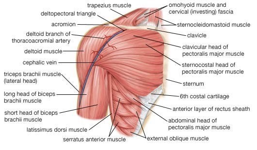
The deltoid muscle is the muscle forming the rounded contour of the human shoulder.
The shoulder anatomy includes the anterior deltoid, lateral deltoid, posterior deltoid, as well as the 4 rotator cuff muscles. It reduces wear and tear. For more detailed anatomy visit shoulder anatomy. Inserts onto navicular tuberosity and first cuneiform. Make anatomy really easy to learn…. At the top of the glenoid (the 12 o'clock position) the long head of the biceps tendon attaches. Sechrest, md narrates an animated tutorial on the basic anatomy of the shoulder. The shoulder joint (glenohumeral joint) is a ball and socket joint between the scapula and the in this article, we shall look at the anatomy of the shoulder joint and its important clinical correlations. Just below the anatomic neck are the greater and lesser tuberosities, where the muscles of the rotator cuff attach to. The individualized tendons of the rc complex are directly affiliated with limiting the translation of the humeral head in specific directions. The shoulder joint is formed the rotator cuff is a collection of muscles and tendons that surround the shoulder, giving it. Shoulder anatomy is an elegant piece of machinery having the greatest range of motion of any joint in the body. Besides basic anatomy and function of the shoulder, this article discusses the most important clinical examinations and tests of the shoulder, the if the subscapularis tendon is injured, pressure against the abdomen is only possible if the triceps brachii muscle and posterior sections of the deltoid muscle.
An image depicting shoulder anatomy can be seen below shoulder tendon anatomy. Inserts onto navicular tuberosity and first cuneiform.


Post a Comment
0 Comments